| |
| S1 |
S2 |
S3 |
 |
 |
 |
 4.4 MB 4.4 MB |
18.2 MB |
13.7 MB |
| 2.2 MB |
10.9 MB |
7.9 MB |
| |
|
|
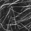
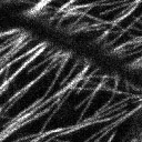
| Microtubule origin, bundle formation, and cortical attachment. Time-lapsed images of Arabidopsis hypocotyl cells expressing GFP-tubulin. Evidence for the initiation of microtubules at the cell cortex (O), the formation of a microtubule bundle (B), and detachment of a microtubule from the cell cortex (D) are illustrated in each cell. Examples of nearby microtubules polymerizing in opposite directions, within array are shown in Movie S3 (AP). The images in each sequence were acquired every 3.8 seconds. Movie 1 consists of 60 frames and Movies 2 and 3 consist of 100 frames each. |
| |
|
|
| S4 |
|
|
 |
|
|
| 4.1 MB |
|
|
| |
|
|
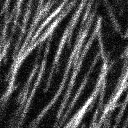
| Evidence for polymer treadmilling. Photobleaching of a line across the cortical microtubule array reveals that both single microtubules and microtubule bundles move by treadmilling (33 images acquired at 8 second intervals). Photobleaching was accomplished using 4 laser scans at 100% laser power. |
| |
|
|
| S5 |
|
|
 |
|
|
| 3.6 MB |
|
|
| 2.0 MB |
|
|
| |
|
|
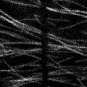
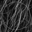
| Measurement of array dynamics by FRAP. Fluorescence recovery after photobleaching (FRAP) experiments were performed using 100% laser power for 4scans in ~10µm diameter circle. 20 images were acquired at 9 second intervals. The green circle denotes the position of the bleached area. Note the recovery of fluorescence in the bleached region shows no obvious spatial bias. |
|
|