|
|
|
|
|
|
|
|
|
|
 |
|
|
|
|
|
|
|
|
|
Epidermal cell pattern |
|
|
|
|
|
|
 |
|
|
|
|
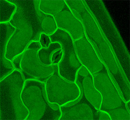
Projection of a confocal image stack showing the edge of a cotyledon. Several cell types are evident; two pairs of guard cells near the center, puzzle piece-like pavement cells, and elongated cells at and near the cotyledon margin. The upper surfaces of cells in the hypodermal cell layer are seen as uniform rafts of signal just below the epidermal cells. This plant is expressing tag Q8, an inframe fusion to PIP2A, a plasma membarne water channel. |
|
|
 |
|
|
|
|
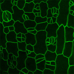
Reconstruction of cotyledon epidermis at an earlier stage of maturation as compared to the image above. Several stages of stomate developement are evident. Tag 29-1. |
|
|
 |
|
|
|
|
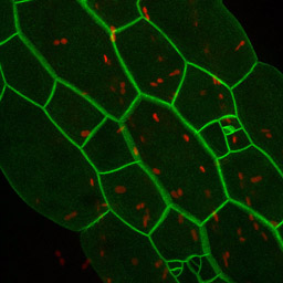
Color merge reconstruction of hypocotyl epidermal cells expressing tag 37-26. The red channel shows plastid autofluoresence. Cell division patterns leading to stomate formation are seen in two cell files. The cells in these two files are shorter in length, and bulge out less from the epidermal surface, than do cells in the adjoining files. |
|
|
 |
|
|
|
|
|
|
Hypocotyl cells expressing tag 5-15. On the left is a 3D rotation movie of the epidermal cell layer. On the right is the original confocal image stack. |
|
|
 |
|
|
|
|
|
|
|
|
|
|
 |
|
|
|
|
|
|