| Embryo structure | |||||||||||||||||||||||||||||
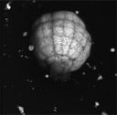 |
Live globular stage embryo expressing soluble EGFP. The embryo was removed from the ovule by cuting the ovule wall and applying gentle pressure. A remanat of the suspensor remins behind at the basal end.
Note the striking symmetry of the embryo and the evident history of cell division patterns, as seen in the relative thickness of the cells walls, leading up to this stage of development. Rotating 3D computer projection. |
||||||||||||||||||||||||||||
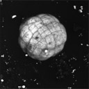 |
Another angle of the same embryo shown above. Note the slight asymmetry at the embronic apex.
Rotating 3D computer projection. |
||||||||||||||||||||||||||||
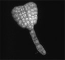 |
Early heart stage embryo expressing soluble EGFP.
Rotating 3D computer projection. |
||||||||||||||||||||||||||||
|
|||||||||||||||||||||||||||||