 |
 |
|
|
| Plant Cell Subcellular Dynamics and 3D imaging Gallery |
 |
 |
 |
 |
 |
 |
 |
 |
 |
Subcellular Dynamics One of the significant advantages of using GFP fusion proteins to label subcellular structure is that dynamic processes can be viewed in live cells with time lapse imaging. On this page we have assembled a sample of movies which highlight the dynamic behavour of plant cells. More images and movies can be found under Cell Structure Tags, Cytoskeletal Dynamics, Cellulose Synthase and Cytokinesis. |
||
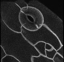
| Peroxisomes | ||
 |
Variable morphology and motility | |
 |
Interconversion of forms | |
 |
Tubule extension and retraction | |
 |
Clustering and tubule anchoring | |
| Endoplasmic reticulum | ||
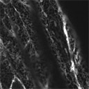 |
Hypocotyl cells | |
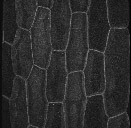
| Vacuole membrane | ||
 |
Hypocotyl cells | |
 |
Complex morphology in trichomes | |
 |
Vacuolar membrane surface | |
| Cytosol | ||
 |
Cytosol dynamics in hypocotyl cells | |
| Peripheral cytosol organization and dynamics | ||
3D Imaging Targeted GFP enables visualization of three dimensional cell and tissue morphology in live organisms. These techniques are useful for the analysis of developmental mutations and for manipulative experiments such as laser ablation. Studies of embryonic development in particular will benefit from these imaging techniques, as the live Arabidopsis embryo has been especially challenging to visualize by traditional methods. |
||
 |
Root | |
 |
Hypocotyl | |
 |
Guard cells | |
 |
Embryos | |