| Hypocotyl cell structure | ||||||||||||||||||||||||||||||||||
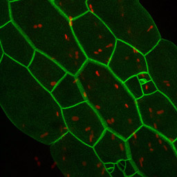 |
Color merge reconstruction of hypocotyl epidermal cells expressing tag 37-26. The red channel shows plastid autofluoresence. Cell division patterns leading to stomate formation are seen in two cell files. The cells in these two files are shorter in length, and bulge out less from the epidermal surface, than do cells in the adjoining files. | |||||||||||||||||||||||||||||||||
 |
 |
Hypocotyl cells expressing tag 5-15. On the left is a 3D rotation movie of the epidermal cell layer. On the right is the original confocal image stack. | ||||||||||||||||||||||||||||||||
 |
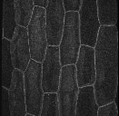
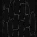
Lower magnification views of hypocotyl cells expressing marker D41. These images highlight the morphology of alternating cell files in the epidermis of the hypocotyl.
3D confocal rotating projection. |
|||||||||||||||||||||||||||||||||
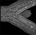 |
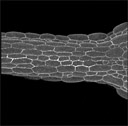
Same plant as shown above, but images include the base of the petioles.
The structure at the crux of the petioles in the image at right is the primordium of the first leaf. A single trichome is evident near the primordium tip. 3D confocal rotating projection. |
|||||||||||||||||||||||||||||||||
|
||||||||||||||||||||||||||||||||||