|
|
|
|
|
|
|
|
|
|
 |
|
|
|
|
|
|
|
|
|
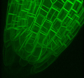
3D root tip cellular anatomy |
|
|
|
|
 |
|
|
|
|
|
 |
|
|
|
|
|
|
|
|
 |
|
|
|
|
Three-dimensional projection of an Arabidopsis root tip expressing tag 37-26. Individual cells can be discerned well into the interior of the root. At extreme angles of rotation fine striations are evident. These striations are the individual confocal planes that make up the reconstructed image, they do not reperesent a biological feature. |
|
|
|
 |
|
|
|
|
|
|
|
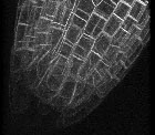
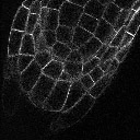
Original confocal image stack. The intensity of fluorescence diminishes as the microscope is focused deeper into the tissue. This is caused by at least three mechanisms. (1) Shading of deeper features by overlying GFP and other light absorbing molecules. (2) Increased scattering of emmitted light by the overlying tissue. (3) Increased optical abberation as the lens is further away from the coverslip. |
|
|
|
|
|
|
|
|
|
|
 |
|
|
|
|
|
|
|
|
|