|
Untagged GFP
|
||||||||||||||||||||||||||||||||||||
 |
||||||||||||||||||||||||||||||||||||
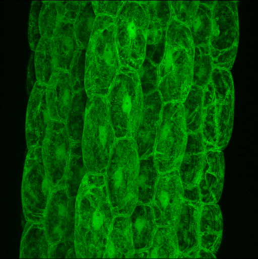
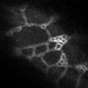
| These images show the distribution of GFP in Arabidopsis cells when no localization tag is present. This pattern formed the baseline for the localization tag screen. GFP is localized to the cytosol and to the nuclear lumen. Because most plant cells are highly vacuolate, the cytosolic localization pattern is surprisingly complicated. GFP patterns change rapidly as cytoplasm is redistributed by cytoplasmic streaming. | ||||||||||||||||||||||||||||||||||||
 |
||||||||||||||||||||||||||||||||||||
 |
 |
|||||||||||||||||||||||||||||||||||
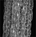
| Hypocotyl cells expressing untagged EGFP from the CaMV 35S promoter. Brightest point projection of a confocal image stack. GFP is localized to the cytosol and the nuclear lumen. These cells are highly vacuolate, confining the cystsol, and thus GFP, to a thin layer at the periphery of the cell and to transvacuolar strands. GFP is largely excluded from the nucleolus, visible as a dark spherical shadow in each nucleus. The movie at left is an animation of the projection shown above, rotated through 60 degrees. The movie at right is the original confocal image stack. The distribution of cytoplasm within each cell is particulary clear in the original image stack. | ||||||||||||||||||||||||||||||||||||
 |
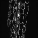
Time series at a single confocal plane of the upper hypocotyl. A variety of dynamic transvacuolar strands are evident. Several nuclei are seen to be deforming and changing position. The images wre aquired at 4 frames a second and are played back at 10 frames per second. | |||||||||||||||||||||||||||||||||||
 |
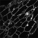
Time series aquired at a plane that just grazes the surface of a petiole epidermal cell. A network of cytsolic strand organization is visible. Strand extension and fusion is visible within the network. | |||||||||||||||||||||||||||||||||||
|
||||||||||||||||||||||||||||||||||||