 |
Chromosomes
|
|||||||||||||||||||||||||||||||||||||
 |
||||||||||||||||||||||||||||||||||||||
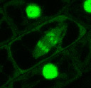
| M253: Strong nuclear accumulation of GFP is observed in interphase cells. In cells undergoing mitosis, the fusion protein highlights condensed chromosomes. In addition to chromosomal association of the GFP tag, there is a higher level of cytosolic fluorescence in cells which display mitotic figures, coincident with the breakdown of the nuclear envelope. The intensity of nuclear labeling is variable from cell to cell, this variability is especially marked in the young root apex.
The tag in this line is the carboxy terminus of Cry2, a blue light photoreceptor. While Cry2 protein has been shown to localize to the nucleus, it remains to be determined if native Cry2 also associates with condensed chromosomes. |
||||||||||||||||||||||||||||||||||||||
 |
||||||||||||||||||||||||||||||||||||||
| Chromosomal association during mitosis | ||||||||||||||||||||||||||||||||||||||
 |
||||||||||||||||||||||||||||||||||||||
| Cell to cell variation | ||||||||||||||||||||||||||||||||||||||
| 180898C: This line shows only moderate accumulation of lable in the nuclei of interphase cells. As nuclei enter mitosis, nuclear fluorescence redistributes to puncta within the nucleus. When the envelope breaks down, a good deal of fluorescence dissapates to the cytosol, but what is left condenses to the location formerly at the center of the nucleus. As cytokinesis proceeds towards metaphase, individual chomosomes can be resolved.
This marker is more uniformly accumulated among cells than is M253. It makes a very nice marker for identifying mitotic cells in the root meristem. |
||||||||||||||||||||||||||||||||||||||
|
||||||||||||||||||||||||||||||||||||||