 |
Tiny bubbles
|
|||||||||||||||||||||||||||||||||||||||
 |
||||||||||||||||||||||||||||||||||||||||
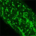
| Markers in this class illumimnate the exterior of a bubble shaped, motile organelle. The identity of this structure has yet to be determined. In addition to the bubble-like structures seen with these markers, the ER membrane is also faintly marked. The movement of the bubbles appears to be correlated with the background ER fluorescence, suggesting they are ER associated. | ||||||||||||||||||||||||||||||||||||||||
 |
||||||||||||||||||||||||||||||||||||||||
| Tiny bubbles in hypocotyl epidermal cells. | ||||||||||||||||||||||||||||||||||||||||
 |
||||||||||||||||||||||||||||||||||||||||
| Tiny bubbles in root cells. | ||||||||||||||||||||||||||||||||||||||||
 |
||||||||||||||||||||||||||||||||||||||||
| Tiny bubbles time series in epidermal cells. | ||||||||||||||||||||||||||||||||||||||||
|
||||||||||||||||||||||||||||||||||||||||